Picture-perfect Proteins
Fall 2021 | By Bree (Adamson) Watson ’04
To make a big impact in the world of bioimaging, it’s wise to look at some of the smallest things in a new light. For Kyu Young Han, assistant professor of optics and photonics, that means developing a new technique to view proteins in human cells. He is creating an innovative tagging system and is building a faster, more efficient super-resolution microscope.
With three recent awards from the National Institutes of Health totaling nearly $3.3 million, Han plans to revolutionize molecular imaging and build an essential microscope for the 4D Nucleome Program, one of the largest collaborative research initiatives since the Human Genome Project was completed in 2003. By developing new technologies to better understand cell functions, Han’s projects have the potential to accelerate researchers’ work with identifying the real-time impact of proteins, which may lead to major advancements in treating diseases.
“I’m excited to be able to contribute my imaging work to this consortium,” Han says. “Our combined contributions highlight the importance of working together to advance understanding of diseases.”
Why Study Proteins?
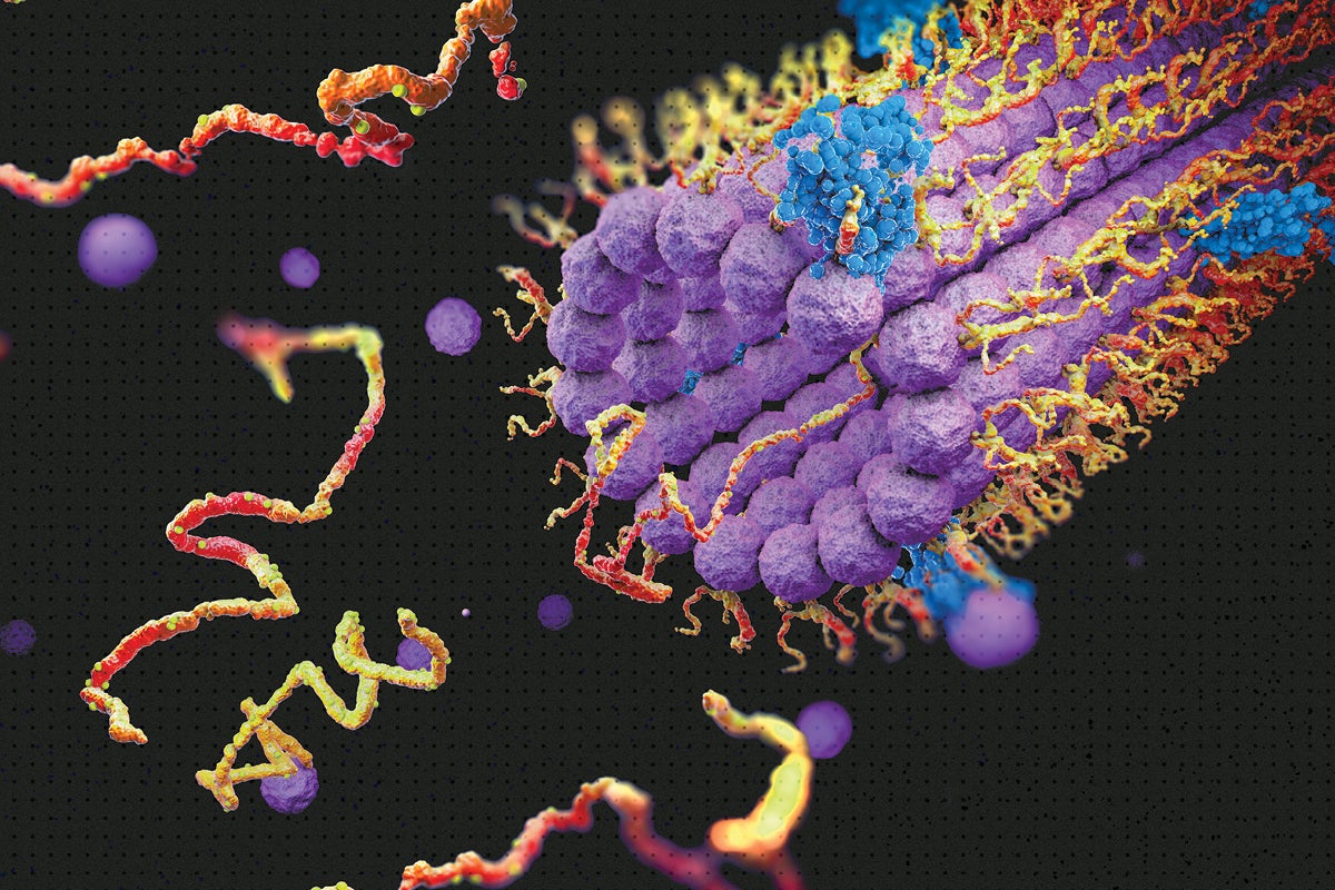
There are an estimated 80,000 to 400,000 proteins in the human body and each one has a specific job, including regulating glucose, moving iron and building muscle. Antibodies, enzymes and transport proteins are some of the protein types that are hard at work in our cells.
Many of these proteins work together to perform programmed functions, but sometimes these interactions deviate from the healthy and expected course of action, which can cause diseases such as cancer, diabetes, and Alzheimer’s. Han’s work aims to help researchers clearly see protein networks and dynamics, which can prevent or improve the negative impacts of certain diseases. Developing technologies to understand how cellular functions in the nucleus affect health and disease is the goal of the 4D Nucleome Program — but Han’s efforts may benefit other major bioimaging initiatives too.
“Scientists realize the genome sequence is not enough information to figure out what causes diseases in the human body,” Han says. “Research consortiums are the next step in diagnosing and treating disease.”
What Makes this Microscope Different?
There are two main types of imaging microscopes — live cell and super-resolution. Live cell microscopy is a minimally invasive method that keeps cells intact but produces images with less detail. Super-resolution microscopes produce high-quality images but can take days or weeks to do so. Han’s new microscope will seamlessly switch between the two imaging styles without losing alignment, gaining crucial insight into protein movement.
“One of our lab efforts is to completely change the engineering, optics and physics of one of the most recent state-of-the-art microscope systems,” Han says. “We are developing a microscopy instrument that produces protein images more accurately and more quickly, and will be maintenance-free, which will make the research process more efficient and less expensive.”
How will proteins be tracked and mapped?
Proteins are always on the move. Researchers can’t rely on just one snapshot of a protein to understand its impact; They need an image with more dimension. That’s why they need a microscope that quickly and accurately captures the locations of proteins and how, when and where they move.
Han and his team of graduate students are also working on a method to engineer antibodies to attach a bar code to target proteins. This innovative technique will allow researchers to identify countless proteins as they interact.
“The microscope we’re developing combined with the antibody bar code tagging technique will enable us to image proteins with more precise resolution at 100 times the speed,” Han says.
How do interdisciplinary interests drive these innovative approaches?
During Han’s undergraduate studies in South Korea, he studied biology for two years before switching to chemistry. While earning his chemistry doctorate, he became interested in optics and photonics after listening to a presentation on super-resolution microscopy, and ever since then he’s built his career on the subject.
“I’m quite lucky to have been exposed to these different areas, and this is why I like bridging several fields of science,” Han says. “I’ve reached out to medical researchers to understand what technical challenges they are faced with. Now I’m trying to provide a new methodology using the speed and precision of optics and photonics.”

Kyu Young Han, Assistant Professor of Optics and Photonics