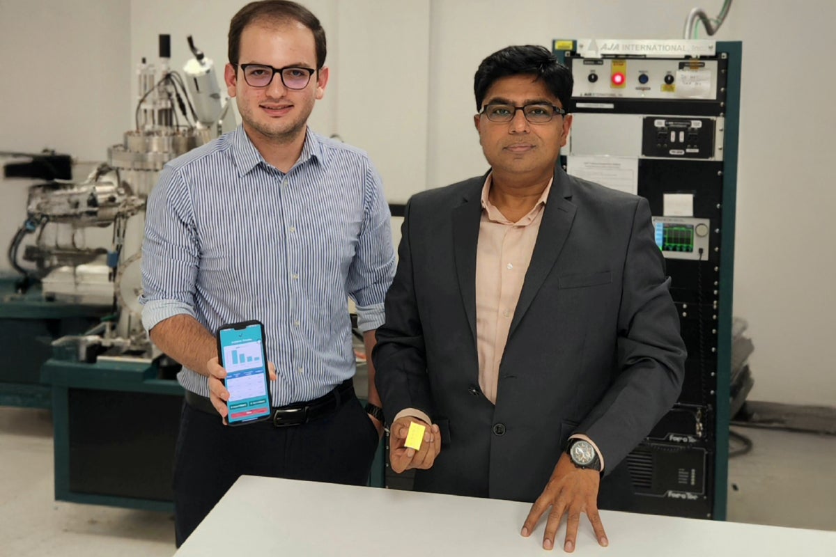
Professor Debashis Chanda (right), working with researchers including physics doctoral student Mahdi Soudi (left), created a smartphone-based biosensor to improve infectious disease detection. The platform provides a faster, more affordable option for communities with limited access to traditional diagnostics.
Early diagnosis of infectious disease is key to slowing outbreaks and improving treatment outcomes. However, current diagnostic techniques are time-consuming, require specialized equipment and are dependent on trained personnel, which hinders accessibility in resource-limited areas.
A Low-Cost, Smartphone-Enabled Diagnostic Platform
UCF researcher Debashis Chanda, a professor at UCF’s NanoScience Technology Center, has developed a self-assembled colorimetric biosensor that can be read using a regular smartphone. The cost-effective platform delivers sensitive and robust detection without needing any sophisticated equipment. The research was recently published and featured as a cover article in Nano Letters, an esteemed scholarly journal published by the American Chemical Society.
Additional researchers on this study include Mahdi Soudi — a physics doctoral student and the lead author of the publication — as well as Caitlin Beech, Pablo Cencillo-Abad, Ishani Chanda, Ángel David Torres Palencia, Amir Ghazizadeh, Pamela Mastranzo-Ortega, Freya Mehta, Javier Sanchez-Mondragón and Abraham Vázquez-Guardado.
The technology combines several novel features:
- Wafer-level fabrication without complex lithography
- Label-free assay format
- Smartphone-enabled readout for portable and low-cost analysis
- Broad dynamic range that spans physiologically relevant IgG concentrations with high reproducibility
Together, these attributes distinguish this approach from earlier colorimetric sensors and showcase its strong potential for real-world applications.
“The sensor works well because of its simple design: a layer of aluminum nanoparticles on a thin optical cavity. This setup makes it very sensitive to small molecular interactions. The sensor uses structural color — like the vivid colors seen in some species — created by the arrangement of two colorless materials. The color can change based on shifts in the local refractive index caused by molecular binding, which alters the resonance and the color seen on the surface. These color changes can be measured using a smartphone,” Chanda says.
Inspired by Vivid Colors
Based on such bio-inspirations, Chanda’s research group innovated a colorimetric sensor, which utilizes the nanoscale structural arrangement of colorless materials to create colors and corresponding changes in colors to sense molecules.
While pigment colorants control light absorption based on the material’s electronic properties — meaning every color needs a new molecule and isn’t sensitive to the surrounding environment — structural colorants control the way light is reflected, scattered or absorbed based purely on the geometrical arrangement of nanostructures and are sensitive to change of medium.
Such structural color-based sensors are environmentally friendly, relying only on metals and oxides, unlike other sensors that use artificially synthesized colorants made from complex, toxic molecules.
Designed for Real-World Use
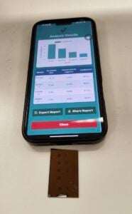
To demonstrate its translational potential, the research team also developed a smartphone application that processes user-captured sensor images and estimates analyte concentration, eliminating the need for bulky optics, spectrometers or trained personnel. This biosensing strategy paves the way for low-cost, rapid, user-friendly diagnostics, empowering individuals to combat infectious diseases and outbreaks more effectively.
“This work introduces a novel platform that addresses the limitations of conventional diagnostic techniques such as complexity, the need for specialized equipment and lack of accessibility,” Chanda says. “Here, we’re not limited by such stringent resource requirements. A smartphone can be used as a diagnostic tool for most point-of-care needs.”
In addition to its diagnostic utility, the platform is highly scalable. More than 20 independent deposition runs supporting over 50 assays showed consistent sensor performance, with yields above 90% and defects mainly due to handling rather than fabrication variability. Because the fabrication relies on thin-film deposition and self-assembly instead of costly lithography, the sensors are inexpensive to produce and compatible with wafer-scale production, making them ideal for disposable point-of-care diagnostics.
Future Research
Chanda says the next steps of the project include further exploration of sensor sensitivity and selectivity aspects to improve its viability as a commercial biochemical sensing platform.
“This biosensing platform holds promise for addressing unmet needs in precise, rapid antibody detection and represents a significant step toward the development of robust, field-deployable biosensors capable of meeting diagnostic requirements in resource-limited and decentralized healthcare environments,” Chanda says.
Licensing Opportunity
For more information about licensing this technology, visit UCF’s Office of Technology Transfer.
Researcher Credentials
Chanda holds joint appointments in UCF’s NanoScience Technology Center, the Department of Physics and the College of Optics and Photonics. He received his doctoral degree in photonics from the University of Toronto and completed a postdoctoral fellowship at the University of Illinois at Urbana-Champaign. He joined UCF in Fall 2012.